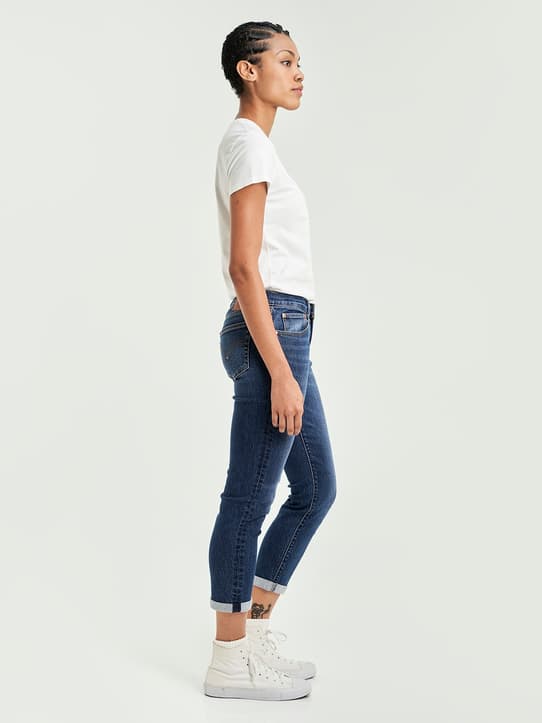
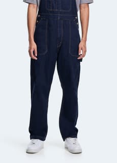
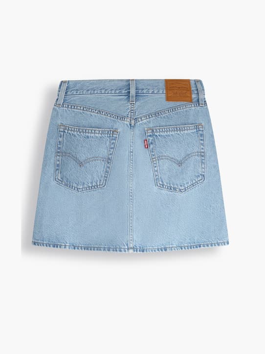
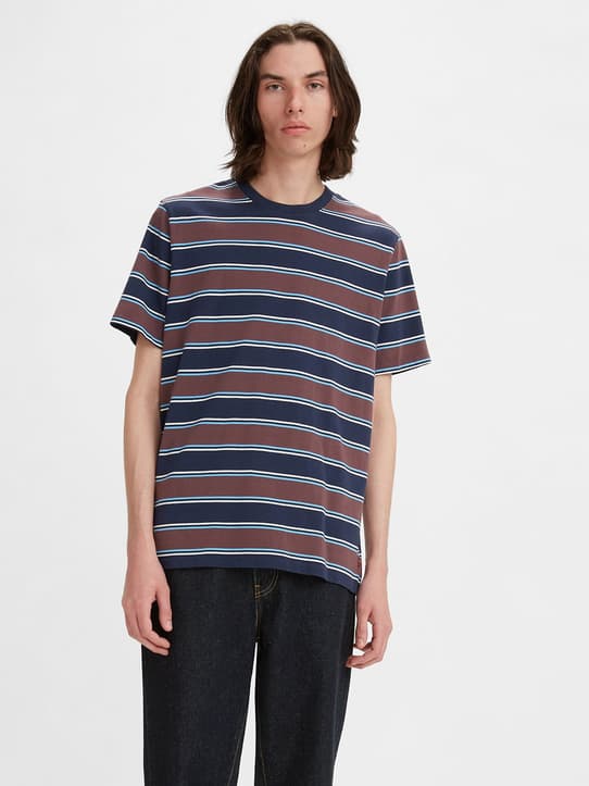
Intercellular transfer of mitochondrial DNA carrying metastasis-enhancing pathogenic mutations from high- to low-metastatic tumor cells and stromal cells via extracellular vesicles | BMC Molecular and Cell Biology

Reagents

GW4869, a neutral sphingomyelinase inhibitor, and tipifarnib, a farnesyl transferase inhibitor, were obtained from Cayman Chemical (Ann Arbor, MI, USA) and ChemScene LLC (Monmouth, NJ, USA), respectively. MitoTracker Red CMXRos, MitoTracker Deep Red FM (MTDR) and CellLight mitochondria-GFP (mtGFP) were supplied by Thermo Fisher Scientific (Waltham, MA, USA). MitoBright LT Red (MitoB LT Red) was purchased from Dojindo Co., Ltd. (Kumamoto, Japan).

Cells and cell culture

High-metastatic A11 cells carrying the ND6 G13997A mutation and low-metastatic P29 cells harboring wild-type mtDNA were established from Lewis lung carcinoma [1, 2, 10]. P29 cells expressing EGFP (EGFP-P29) were established by introduction of the pEGFP-N1 expression plasmid followed by G418 selection and cloning. WA-mFib cells, a mouse stromal cell line for human lung small cell carcinoma, and mouse RAW264.7 macrophages (RCB1925 and RCB0535, respectively, from RIKEN BioResource Research Center) [38] were cultured in Dulbecco’s modified Eagle’s medium (DMEM) supplemented with 10% FBS and 40 μg/ml gentamicin in a humidified atmosphere with 95% air/5% CO2 at 37 °C. CTLL-2 cells, a mouse cytotoxic T-cell line (RCB0637, RIKEN BioResource Research Center), were cultured in DMEM supplemented with 10% FBS and 50 ng/ml recombinant mouse IL-2 (BioLegend, San Diego, CA, USA). Human cervical cancer HeLa cells, human lung carcinoma A549 cells, human colon carcinoma DLD1 cells and human pancreatic carcinoma MIAPaCa-2 cells [39] were cultured in DMEM/10% FBS. MtDNA-less P29 (ρ0P29) cells [1] and ρ0HeLa cells (EB8 cells) [40] were cultured in DMEM supplemented with 0.1 mg/ml sodium pyruvate and 0.5 mg/ml uridine. Mouse embryonic fibroblasts (MEFs) were prepared and cultivated as described previously [41]. The addition of EVs to ρ0 P29 cells and ρ0HeLa cells was performed in the presence or absence of the EV-Entry System (System Biosciences, Palo Alto, CA, USA), which increases the rate of EV uptake and offloading of the cargo into the cytoplasm of the recipient cells.

Labeling of mitochondria

For in vitro experiments, the cells and EVs were labeled with MTDR or mtGFP and MitoTracker Red, respectively. For in vivo experiments, cells were stained with MitoB LT Red, which is designed for mitochondrial retention for long-term visualization and shows stronger fluorescence signals than other commercially available dyes (Dojindo). Staining of the cells was performed according to the manufacturer’s instructions. The cells were observed under a Leica TSC SP8 confocal laser scanning microscope (Leica Microsystems, Wetzlar, Germany).

Coculture

A11-MTDR or P29-MTDR cells (5 × 104 cells) and P29-mtGFP, A11-mtGFP cells (5 × 104 cells), respectively, were cocultured on glass coverslips in a well of a 12-well multiwell culture plate for 24 h. In other experiments, A11-MTDR cells (5 × 104 cells) were cocultured with EGFP-P29 cells (5 × 104 cells), WA-mFib-mtGFP cells (5 × 104 cells), RAW264.7-mtGFP cells or MEF-mtGFP (5 × 104 cells) for 24 h. The cells were fixed with 4% paraformaldehyde and observed under a confocal laser microscope. A11-MTDR cells (5 × 104 cells) were also cocultured with CTLL-2-mtGFP cells (1 × 105 cells) in a 35-mm glass-bottom culture dish for 24 h and observed under a confocal laser microscope without fixation.

Preparation of EVs from cell culture media and normal mouse serum

EVs in cell culture medium were isolated by ultracentrifugation [42] with some modifications. Briefly, cells were cultured in DMEM containing 10% exosome-free FBS for 2 or 3 days. Exosome-free FBS was prepared by ultracentrifugation at 110,000×g overnight at 4 °C. The conditioned medium was centrifuged at 400×g for 10 min to remove cells, and the supernatant was centrifuged at 2000×g for 20 min to remove cell debris and apoptotic bodies. The resulting supernatant was ultracentrifuged at 15,000×g for 30 min at 4 °C; the pellet was washed with Dulbecco’s phosphate buffered saline (DPBS) and used as large EVs (L-EVs). The supernatant was passed through a Millex-GP filter (0.22 μm pore) (Merck-Millipore Burlington, MA, USA) and ultracentrifuged at 110,000×g for 90 min at 4 °C. The pellet was washed with DPBS to eliminate contaminated proteins and centrifuged again at 110,000×g for 90 min at 4 °C. The pellet was resuspended in DPBS and used as small EVs (S-EVs); EVs were sterilized by filtration through a Millex-GV filter (0.22 μm pore). EVs in C57BL/6 mouse serum were isolated using Total Exosome Isolation Reagent (from serum) (Thermo Fisher Scientific).

Characterization of isolated S-EVs

The size range of the isolated S-EVs from the culture media of A11 cells was measured by a Zeta Potential/Particle Size Analyzer (Otsuka Electronics, Osaka, Japan). Annexin A1, CD9, CD63, CD81, GAPDH, LC3B, β-actin, Lamin A/C and mitochondrial proteins were detected by Western blot analyses using rabbit polyclonal anti-Annexin A1 antibody (Cell signaling Technology, Danvers, MA, USA), rabbit polyclonal anti-CD9 antibody (Flarebio Biotech LLC, Baltimore, MD, USA), rabbit polyclonal anti-CD63 antibody (Flarebio Biotech LLC), rabbit polyclonal anti-CD81 antibody (Cell signaling Technology), rabbit monoclonal anti-GAPDH antibody (Cell signaling Technology), rabbit polyclonal anti-LC3B antibody (Cell signaling Technology), mouse monoclonal anti-β-actin (Santa Cruz Biotechnology, Dallas, TX, USA), mouse monoclonal anti-Lamin A/C antibody (Santa Cruz Biotechnology) and Membrane Integrity WB Antibody Cocktail (abcam, Cambridge, UK). For this, EVs were dissolved in SDS-sample buffer, and the particle concentration was adjusted based on the protein concentration measured by the BCA method using bovine serum albumin (BSA) as a standard. For preparation of cell lysates of P29 and A11 cells, they were lysed in RIPA buffer containing cOmplete Protease Inhibitor Cocktail (Merck, Kenilworth, NJ, USA) and PhosSTOP (Merck Millipore, Billerica, MA, USA). The lysates were centrifuged at 10,000×g for 10 min at 4 °C, and the supernatants were used for immunoblot analysis. A11 mitochondria were isolated by using Mitochondria Isolation Kit for Cultured Cells (abcam). After SDS-polyacrylamide gel electrophoresis, the proteins were transferred to an Immobilon-P transfer membrane (Merck Millipore, Billerica, MA, USA). The membrane was incubated with washed with TBS-T (5 mM Tris/13.8 mM NaCl/0.05% Tween-20), and then incubated with horseradish peroxidase (HRP)-conjugated goat anti-rabbit IgG or goat anti-mouse IgG. Proteins were detected using ECL Plus Western blotting detection reagent (Amersham Biosciences, Piscataway, NJ, USA). For staining of EVs with PKH67, isolated L-EVs and S-EVs were stained using PKH67 Fluorescence Cell Linker Kits (Sigma-Aldrich, St. Louis, MO, USA) for 5 min, resuspended in DPBS, and centrifuged again at 15,000×g for 30 min and 110,000×g for 90 min, respectively, at 4 °C. The pellet was resuspended in DPBS.

Transmission electron microscopy (TEM)

Negative-stain TEM and cryo-TEM of A11 S-EVs were performed by JEOL Ltd. (Tokyo, Japan).

Detection of mtDNA in EVs and ρ0 cells by polymerase chain reaction (PCR)

For detection of mtDNA in EVs, the EVs were dissolved in 20 mM NaOH, incubated for 10 min at 95 °C, neutralized with 1 M Tris-HCl (pH 8.0), and then subjected to PCR analysis. For detection in mtDNA in ρ0P29 cells, the cells were detached from culture dishes with trypsin-EDTA (Sigma-Aldrich, Co., LLC, Saint Louis, MO, USA) at 37 °C for 10 min, washed extensively with DPBS three times, and then dissolved in DirectPCR Lysis Reagent (Cell) (Viagen Biotech, Inc., Los Angeles, CA, USA) containing 0.2 mg/ml proteinase K. After incubation at 55 °C for 60 min with shaking, proteinase K was inactivated by heating at 85 °C for 45 min. PCR was performed using GoTaq Hot Start DNA polymerase (Promega Corp., Madison, WI, USA). The PCR conditions were 94 °C for 5 min followed by 32 cycles of 94 °C for 30 s, 58 °C for 30 s, and 72 °C for 30 s. The primers used were 5′-CCGGGCCCATTAAACTTGGG-3′ and 5′- TAGTGTTTTTGGGGTTTGGCATT-3′ for HVR, 5′-CTAGCAGAAACAAACCGGGC-3′ and 5′-ATGGTGGTACTCCCGCTGTA-3′ for ND1, 5′-GACTTGCAACCCTACACGGA − 3′ and 5′-TGTGGTGTAAGCATCTGGGT-3′ for COI, 5′-ACCACTAACCTGACTATCAAGCC-3′ and 5′-GTTTGGTTCCCTCATCGGGT-3′ for ND4, 5′-GTTGGTTGTCTTGGGTTAGCAT-3″ and 5′-CTACCCCAATCCCTCCTTCCA-3′ for ND6, and 5′-CTCTGGCTCCTAGCACCATGAAGA-3′ and 5′-GTAAAACGCAGCTCAGTAACAGTCCG-3′ for ACTB. Aliquots of the PCR products were electrophoresed in 1.5% agarose gels and visualized on a transilluminator after staining with ethidium bromide. For recognition of the G13997A mutation by PCR-RFLP, a 254 bp fragment containing the 13,997 G > A site was amplified by PCR using the mismatched primers forward primer: 5′-CCCACTAACAATTAAACCTAAACCTCCATActTA-3′, (small letters indicate the mismatch site) and reverse primer: 5′-GGGGCAGGTAGGTCAATGAA-3′ to create a restriction site for AflII [1]. After digestion with AflII, the restriction fragments (223 bp and 31 bp) were separated in a 3% agarose gel. For detection of mtDNA in EVs from human cancer cells, the following primers were used: 5′- TCTTTCATGGGGAAGCAGAT-3′ and 5′- GCACTCTTGTGCGGGATATT-3′ for HVR, 5′- TCATGACCCTTGGCCATAAT-3′ and 5′- GGGGAATGCTGGAGATTGTA-3′ for ND1, 5′- ACGTTGTAGCCCACTTCCAC-3′ and 5′- GGGTTCTTCGAATGTGTGGT-3′ for COI, 5′-TGAACGCAGGCACATACTTC-3′ and 5′-TGTTTGTCGTAGGCAGATG-3′ for ND4, and 5′- CCCCGAGCAATCTCAATTAC-3′ and 5′-GGTGTGGTCGGGTGTGTTAT-3′ for ND6. For the detection of mtDNA in ρ0HeLa cells, the following primers were used: 5′- GTTGGTTGTCTTGGGTTAGCAT-3′ and 5′- CTACCCCAATCCCTCCTTCCA-3′ for ND6, and 5′-TGACGGGGTCACCCACACTGTGCCCATCTA-3′ and 5′- CTAGAAGCATTTGCGGTGGACGATGGAGGG-3′ for ACTB.

Animal experiments

Five- to six-week-old male C57BL/6 J mice (CLEA Japan, Osaka, Japan) were used in this study. The mice were housed in a barrier facility under specific pathogen-free conditions and assessed with regard to health every day. For the in vivo mitochondrial distribution in tumor tissues, syngeneic transplantable P29 cells or EGFP-P29 cells (1 × 106 cells) were subcutaneously implanted into the mice. When the tumor size reached approximately 0.75 cm3, another syngeneic A11 cell line (5 × 106 cells) labeled with MitoB LT Red was injected intratumorally. After 3 days, homograft tumors were excised and immediately embedded and frozen in OCT compound for subsequent analysis. The mice were euthanized by CO2 inhalation at the end of the study.

Immunostaining

Cryostat sections (7 μm thick) of homograft tumor tissues containing EGFP-P29 and MitoB LT Red-labeled A11 cells were fixed with 4% paraformaldehyde and stained with rabbit polyclonal anti-GFP antibody (Proteintech, Sankt Leon-Rot, Germany) followed by Alexa Fluor 488 (AF488)-conjugated anti-rabbit IgG. In this case, to clearly visualize EGFP-P29 cells, immunostaining with anti-GFP antibody was employed. Cryostat sections of tumor tissues containing P29 and MitoB LT Red-labeled A11 cells were fixed with 4% paraformaldehyde for 10 min and blocked with 1% BSA in DPBS. The sections were treated with an M. O. M. Immunodetection Kit (Vector Laboratories, Burlingame, CA, USA) and then incubated with mouse monoclonal anti-α-SMA (clone 1A4) (Dako, Jena, Germany) antibody followed by AF488-conjugated goat anti-mouse IgG. The sections were counterstained with DAPI and observed under a Leica TSC SP8 confocal laser scanning microscope. WA-mFib cells were also immunostained with the anti-α-SMA antibody followed by AF594-conjugated goat anti-mouse IgG.

Read more here: Source link